04/26/2013
Reproduced, with permission, from Ref. 1 © 2013 American Chemical Society
Rubber is an important class of material with applications ranging from high-performance devices to the humble car tire. But characterizing the complex blend of cross-linked polymers and carbon ‘filler’ particles that comprises rubber is far from straightforward. To better investigate its composition, researchers have turned to atomic force microscopy (AFM) — a technique that uses tiny, oscillating silicon probes to measure the physical properties of surfaces.
Conventional AFM instruments are unable to fully investigate the behavior of rubber because their probes cannot vibrate sufficiently fast across different frequencies. Now, Ken Nakajima and colleagues from the AIMR at Tohoku University have solved this problem with a design that uses so-called ‘piezoelectric’ crystal actuators to boost the sensors’ vibrational capabilities1. While actuators allow the three-dimensional scanning of surfaces, they are designed for large movements, meaning that AFM instruments have a limited frequency range. To obviate this issue, the researchers inserted an additional piezoelectric actuator — small, lightweight and with large resonant frequencies — above the AFM scanner to simultaneously oscillate the sample and probe it up to 20,000 times per second.
The team tested its design on a series of isoprene and styrene–butadiene rubbers. First, they determined mechanical contact parameters by recording how increasing force deformed the materials. They then compared the amplitude and phase of AFM signals from the rubbers to those of a solid mica surface. Repeating the measurements at different frequencies generated ‘viscoelastic’ parameters that described the rubbers’ dynamic deformation.
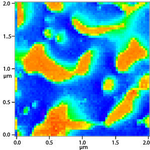
Two-dimensional ‘maps’ of the mechanical loss of a mixed rubber surface were created using the AFM scanning functions (see image). These revealed an ‘island’-like arrangement of nanoscale isoprene rubbery regions surrounded by a ‘sea’ of styrene–butadiene rubbery regions. Critically, the immiscible nature of the rubbers only became clear when scanning at a certain range of frequencies.
Nakajima notes that the ability to measure frequency-dependent deformation can eliminate many of the time-consuming tests currently needed to characterize rubber. “In viscoelastic materials, frequency and temperature are believed to be related. Therefore, measurements with narrow frequency ranges must be performed at multiple temperatures. Our instrument can carry out these measurements with a much wider frequency range at a fixed temperature,” he says.
Furthermore, explains Nakajima, the AFM measurements match many of the frequencies encountered during macroscopic testing. Such direct comparisons of large- and nano-scale phenomena are vital to comprehend multi-component systems. “Our technique can visualize the viscoelastic nature of each component in high resolution, giving valuable feedback for future materials design.”
Igarashi, T., Fujinami, S., Nishi, T., Asao, N. & Nakajima, K. Nanorheological mapping of rubbers by atomic force microscopy. Macromolecules 46, 1916–1922 (2013). | article
This research highlight has been approved by the authors of the original article and all information and data contained within has been provided by said authors.