
08/27/2012

© 2012 X. Shi
Bone health hinges on the assembly of various cells, including osteoblasts and osteoclasts, into well-defined functional structures that manage bone-specific tasks in the body, such as cell growth, differentiation and protein secretion. However, tumor-induced injuries and other bone-related diseases hinder these self-regulated, sophisticated tasks. To address these conditions it is essential to develop tissue engineering approaches that direct cell behavior.
Xuetao Shi and Hongkai Wu and co-workers from the AIMR at Tohoku University, together with colleagues in Hong Kong and Beijing1, have now developed patterns that serve as scaffolds for bone regeneration. The patterns consist of polymer microspheres filled with drug or protein molecules that are regularly interspaced.
Prevalent methods for directing cell behavior exploit either chemical and biological signals or topographical cues, but using each of these methods separately has proven ineffective. The former approach can regulate stem cell differentiation into specialized cells but does not properly control cell spatial arrangements, while the latter faces opposite limitations.
By combining both approaches into microsphere patterns, the researchers have now taken advantage of chemical and physical stimulations at the same time. The patterns induced an aligned growth of cells that simulates the native surfaces of bones and the membrane enveloping their outer surface. “We also used the microspheres to encapsulate and release drug molecules to mimic the in vivo bone environment, which contains various beneficial substances such as growth hormones,” adds Shi.
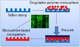
To create the patterns (see image), Shi’s team first mixed an organic solution containing the biocompatible, biodegradable poly(lactic-co-glycolic acid) polymer with a drug or protein dissolved in water in the presence of emulsifiers. After depositing the resulting microspheres onto a patterned Teflon mold and adding a solvent to ensure inter-particle cohesion, the researchers covered the mold with a Teflon slide and applied pressure to this lid to obtain the patterned material.
Initial tests showed that patterns with narrow grooves between the microspheres resulted in better cell alignment than wide ones. Moreover, the microspheres efficiently encapsulated and slowly released hydrophilic and hydrophobic molecules, prompting the researchers to simultaneously load multiple drugs into them. Stem cells cultured on these multi-drug-loaded patterns exhibited high levels of bone tissue formation markers, proof of their enhanced differentiation towards osteoblasts.
In addition to bone repair, this combined chemical–topographical strategy can be used to study muscle and blood vessel cells for potential regenerative therapies of cardiac tissues. “Our future work will focus on further developing scaffolds displaying surfaces that mimic native tissue and drug or gene release function for organ and tissue regeneration,” says Shi.
Shi, X., Chen, S., Zhou, J., Yu, H., Li, L. & Wu, H. Directing osteogenesis of stem cells with drug-laden, polymer-microsphere-based micropatterns generated by Teflon microfluidic chips. Advanced Functional Materials 22, 3799–3807 (2012). | article
This research highlight has been approved by the authors of the original article and all information and data contained within has been provided by said authors.