03/31/2014
© 2014 Chunlin Chen
Over the course of its lifetime, a typical shark will lose and regrow thousands of teeth as they become embedded in prey or worn out. None of these teeth, however, ever suffers from cavities. Recent studies have discovered that the hard, enamel-like coating of a shark tooth is made from fluorapatite — a fluorinated calcium phosphate material that is an active ingredient in most toothpastes. Although researchers already attribute high concentrations of fluoride ions to low levels of tooth decay, direct evidence of this chemical’s cavity-reducing action remained elusive.
Chunlin Chen and colleagues from the AIMR at Tohoku University, in collaboration with researchers from across Japan, have now used transmission electron microscopy (TEM) to capture the first images of individual atoms inside shark teeth1. Their findings show that unusual bonding interactions between fluorine and calcium atoms may be critical to understanding fluorine’s tooth-strengthening capabilities.
For decades, scientists have used TEM to observe the atomic-scale structure of inorganic materials. Shark teeth, however, are biominerals — complex substances containing both delicate organic matter and robust inorganic minerals, which are easily damaged by high-energy electron beams. Current understanding of the enamel-like structures that envelop shark teeth is thus limited to micrometer-sized regions.
To overcome this challenge, the researchers used a scanning transmission electron microscope equipped with special ‘aberration-corrected’ lenses and low-dose imaging capabilities. The approach employs a small lens aperture that disperses the electron beam over a wider area than usual, thereby lowering the beam intensity. However, because such a low-dose beam results in noisy TEM images, the researchers had to search for a careful balance between irradiation-induced damage and image quality.
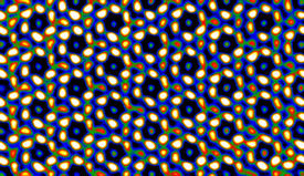
When the team examined a tooth from an Isurus oxyrinchus — also known as a shortfin mako shark — TEM images showed that its enamel-like structure contained bundles of single-crystalline fluorapatite nanorods, roughly fifty nanometers wide. Deeper probing revealed a hexagonal atomic framework inside the nanorods, with calcium, phosphorus and oxygen surrounding a central fluorine atom (see image). This structure suggests that fluorine is critically important to shark tooth enamel as its loss would destabilize the entire chemical structure.
Computer simulations of the structure of fluorapatite further illustrated fluorine’s unique role in dental health. The researchers were able to determine that bonding between fluorine and calcium contained both ionic and covalent contributions, making it stronger than typical fluorine bonds. “The direct imaging of fluorine atoms and its surprising mixed covalent–ionic bonding should be of fundamental significance to the field of dentistry,” says Chen.
Chen, C., Wang, Z., Saito, M., Tohei, T., Takano, Y. & Ikuhara, Y. Fluorine in shark teeth: Its direct atomic-resolution imaging and strengthening function. Angewandte Chemie International Edition 53, 1543–1547 (2014). | article
This research highlight has been approved by the authors of the original article and all information and data contained within has been provided by said authors.