
10/29/2012

© 2012 Royal Society of Chemistry
Natural tissues are highly organized structures, often formed from multiple cell types precisely positioned to carry out their required roles. Efforts to mimic these structures in order to create artificial tissues — for example, to help heal parts of the body that have sustained damage from injury or disease — is no simple task.
Recently developed techniques such as dielectrophoresis use electric fields to position living cells within a three-dimensional matrix; however, trapping the cells in place while ensuring their long-term viability has proven difficult. A highly biocompatible scaffold material that could solve this problem has now been identified by an international team of researchers led by Ali Khademhosseini and Tomokazu Matsue from the AIMR at Tohoku University1.
The researchers selected a semi-natural hydrogel material based on gelatin — gelatin methacrylate (GelMA) — to use as a tissue scaffold. Previous studies have shown that GelMA is a suitable material for culturing cells. The researchers also observed that GelMA had the capability to bypass the typical pitfalls of the most commonly used tissue engineering scaffolds. “Owing to its low viscosity and conductivity, we suspected that the GelMA hydrogel could be a promising candidate for cell manipulation using the dielectrophoresis method,” says Samad Ahadian, a member of the AIMR team.
Testing GelMA in the laboratory, the researchers confirmed that it was a suitable matrix within which to guide cells into position using dielectrophoresis. Once the cells were in place, the team exposed the scaffold to UV light. This triggered a chemical cross-linking reaction within the hydrogel, which forms the polymer matrix and traps the cells in place. Using a photomask, the researchers were able to trap one type of cell in one part of the polymer before introducing and trapping a second cell type within the same scaffold.
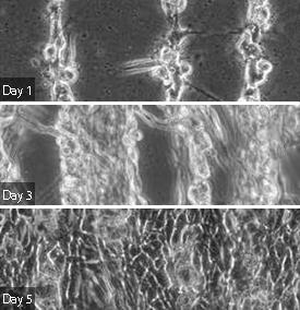
Crucially, the cells retain long-term viability after the formation of the cross-linked polymer, and readily proliferate over several days (see image). The natural gelatin on which the hydrogel is based is responsible for this high biocompatibility. “The GelMA comprises natural cell-binding motifs that support cell adhesion, migration, and proliferation,” explains Ahadian. “The optimized GelMA concentration of 5% ensures that there is plenty of space for the encapsulated cells to obtain the nutrients required and repel wasted products.”
The next step will be to produce engineered tissues with differentiated cell types, from neural to muscle cells. According to Ahadian, the potential applications extend beyond damaged tissue repair to include uses in drug screening models or as bio-actuators.
Ramón-Azcón, J., Ahadian, S., Obregón, R., Camci-Unal, G., Ostrovidov, S., Hosseini, V., Kaji, H., Ino, K., Shiku H., Khademhosseini, A. & Matsue, T. Gelatin methacrylate as a promising hydrogel for 3D microscale organization and proliferation of dielectrophoretically patterned cells. Lab on a Chip 12, 2959–2969 (2012). | article
This research highlight has been approved by the authors of the original article and all information and data contained within has been provided by said authors.