
08/27/2012

© 2012 Wiley-VCH Verlag GmbH & Co. KGaA, Weinheim
Traditional methods of culturing cells for biological and biomedical applications use two-dimensional cell cultures, typically growing cells on the base of plastic Petri dishes. In recent years, however, a three-dimensional (3D) environment has emerged as more appropriate for cell culture as it replicates the biological milieu. This is particularly important for embryonic stem cells which can grow into a variety of different tissue structures (such as bone, nerve, cartilage cells) from 3D embryoid bodies. Researchers from the AIMR and the Graduate School of Environmental Studies at Tohoku University have now built an electrochemical device that monitors the activity and differentiation of stem cells in an embryoid body1.
Usually, scientists label biomolecules for analysis with fluorescent markers to identify and quantify their presence. But labelling molecules may be toxic or interfere with cellular behavior, and fluorescence levels can be compromised by autofluorescence (signals from unlabeled molecules) or quenching from other non-target materials or liquid turbidity. The electrochemical device developed by Tomokazu Matsue, Kosuke Ino and colleagues does not require these labels, thus removing these problems.
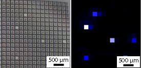
Electrochemical detection has been used before. However, this device has a unique feature. Detection is achieved using an array of 256 electrochemical sensors with only 32 bonding pads for external connection (see image), placed at the base of deep microwells which enables spatially-resolved measurements. “This electrochemical sensor density is the highest in the field of electrochemical lab-on-a-chip devices,” the researchers explain.
The research team quantified cellular activity from embryoid bodies on the array by ‘redox cycling,’ whereby alkaline phosphatase activity (a marker for the differentiation level of embryonic stem cells) was measured by the reduction and oxidation of one of its reaction products. Potentials of electrodes in each sensor were set at levels to initiate redox cycling at a specific location from which the local current signals were collected and measured.
Interestingly, enzymatic activity as measured by the electrode device after a four-day incubation did not increase as expected, suggesting that the stem cells had differentiated. The device will therefore be useful to screen embryoid bodies’ differentiation levels, and will provide more quantitative than conventional sorting by morphological analysis.
Furthermore an embryoid body resting on top of the sensor array was imaged without cross-talk from molecular diffusion between sensors due to the microwells. In the next stage of this project, the researchers will explore the limits of this imaging capability. “We intend to increase the density of the electrochemical sensors even further for electrochemical imaging of a single cell or a single tissue,” say the researchers.
Ino, K., Nishijo, T., Arai, T., Kanno, Y., Takahashi, Y., Shiku, H. & Matsue, T. Local redox-cycling-based electrochemical chip device with deep microwells for evaluation of embryoid bodies. Angewandte Chemie International Edition 51, 6648–6652 (2012). | article
This research highlight has been approved by the authors of the original article and all information and data contained within has been provided by said authors.