
07/30/2012

© 2012 PNAS
Electroactive and short-lived species that are released and consumed by cells, including neurotransmitters and reactive oxygen-based molecules, are central to cell metabolism, but their detection at cell surfaces and interfaces remains challenging. A research team led by Tomokazu Matsue and Yasufumi Takahashi from the AIMR1, in collaboration with Yuri Korchev from Imperial College London, has now developed a high-resolution, non-invasive imaging method called voltage-switching mode–scanning electrochemical microscopy (VSM–SECM). The technique, which can be utilized under physiological conditions, provides high-quality topographical and electrochemical images of living cells simultaneously.
Many SECM imaging approaches have been developed to determine surface topology and reactivity. However, they usually measure interactive forces by direct contact, through the tip of a tiny electrode that moves across the surface of the substrate being imaged — which can damage cell membranes.
Matsue and colleagues have now used faradaic current, which is generated by the reacting electroactive species, to control the motion of the electrode, and continuously prevent it from touching the substrate surface. Moreover, they fabricated nanometer-sized glass-insulated carbon electrodes that allow for high-resolution imaging.
To initiate one measurement, the team moved the electrode tip towards the surface while monitoring the distance-dependent current created by the hindered diffusion of the electroactive species as they underwent reduction. When the tip current decreased to a level that reflected the desired distance between probe and either the substrate surface or an electroactive molecule, the researchers stopped the probe and recorded its position. At each position, the team switched the voltage applied to the electrode to also determine the species’ activity. Repeating this process at several points across the surface yielded simultaneous topographical and electrochemical images.
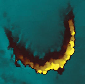
Similar to ultra-high resolution images, the resulting maps highlighted the small protrusions and wave structures of skin and cardiac cells as well as elongated hair cells (see image). Visualization of cell membrane proteins associated with cancer showed that the protein distribution was uneven and did not match the ultra small features observed from the topological images — demonstrating the usefulness of VSM-SECM for chemical mapping at biological interfaces. Simultaneous topological and electrochemical measurements have also enabled Matsue’s team to detect the release of neurotransmitters from neurons. Furthermore, the combination of SECM with fluorescence microscopy clearly showed the extremities of neurons at the synapse.
Currently, the researchers are further exploring neuronal communication using their new method. “We would like to map the neurotransmitter releasing sites,” says Matsue. The team is also planning to monitor the release-related changes in neuron topography.
Takahashi, Y., Shevchuk, A. I., Novak, P., Babakinejad, B., Macpherson, J., Unwin, P. R., Shiku, H., Gorelik, J., Klenerman, D., Korchev, Y. E. & Matsue, T. Topographical and electrochemical nanoscale imaging of living cells using voltage-switching mode scanning electrochemical microscopy. Proceedings of the National Academy of Sciences USA 109, 11540–11545 (2012). | article
This research highlight has been approved by the authors of the original article and all information and data contained within has been provided by said authors.